how accurate is fetal anatomy ultrasound Transvaginal ultrasound requires covering the ultrasound transducer in a plastic or latex sheath which may cause a reaction in women with a latex allergy. Snijders RJ Shawa L Nicolaides KH.
How Accurate Is Fetal Anatomy Ultrasound, You might have heard of the baby heartbeat testit involves analyzing the speed of the fetal heartbeat in order to determine whether the baby is a boy or a girl. Aetna considers in-office and in-hospital antepartum fetal surveillance with non-stress tests NST contraction stress tests CST biophysical profile BPP modified BPP and umbilical artery and middle cerebral Doppler velocimetry medically necessary according to the American College of Obstetricians and Gynecologists ACOG Clinical Guideline on. It is very likely that you will get a glimpse of what are now very obvious boy parts during this type of ultrasound.
 Nub Theory For Detecting Gender As Early As 10 Weeks Baby Gender Predictor Baby Gender Prediction Baby Gender Ultrasound From pinterest.com
Accurate performance of an ultrasound. Recent applications of deep learning in medical US analysis have involved various tasks such as traditional diagnosis tasks including classification segmentation detection registration biometric measurements and quality assessment as well as emerging tasks including image-guided interventions and therapy Of these classification detection and. CRL is measured as the largest dimension of embryo excluding the yolk sac and extremities.
Ultrasound images are captured in real-time so they can show the structure and movement of internal organs as well as blood flowing through blood vessels.
Recent applications of deep learning in medical US analysis have involved various tasks such as traditional diagnosis tasks including classification segmentation detection registration biometric measurements and quality assessment as well as emerging tasks including image-guided interventions and therapy Of these classification detection and. The major fetal anomalies that can be recognized by ultrasound in the first trimester. Assessment of risk based on ultrasound findings and maternal age. An NT is a special type of ultrasound using a very sensitive but safe machine. Predominantly fetal growth and organ maturation23.
Another Article :

If you have chosen to find out the sex of your baby you are most likely going to do so via ultrasound. For an accurate measurement the fetal head should be in the transverse plane. Historically the second trimester ultrasound was often the only routine scan offered in a pregnancy and so was expected to provide information about gestational age correcting menstrual dates if necessary fetal number and type of multiple pregnancy. An NT is a special type of ultrasound using a very sensitive but safe machine. Most of the time the condition is suspected during a physical exam following birth. Pin On Radiologymasterclass.

Typically the ultrasound is done halfway through the pregnancy. By an accurate early scan subsequent scans should not be used to recalculate the gestational age1. Correct evaluation depends on the accuracy of the gestational age being used the precision of the weight measurements and using a weight curve that represents the population being studied. Fetal Pole 55-6 weeks The fetal pole or developing embryo should be seen at 55-6 weeks gestational age by transvaginal ultrasound. In these Guidelines we assume that the gestational age is known and has been determined as described above the pregnancy is singleton and the fetal anatomy is normal. Accuracy Of Fetal Gender Determination In The First Trimester Using Three Dimensional Ultrasound Youssef 2011 Ultrasound In Obstetrics Amp Gynecology Wiley Online Library.

For an accurate measurement the fetal head should be in the transverse plane. Most of the time the condition is suspected during a physical exam following birth. Ultrasound images are captured in real-time so they can show the structure and movement of internal organs as well as blood flowing through blood vessels. Congenital hydrocephalus may be diagnosed by fetal ultrasound. The MCA vessels are often found with color or power Doppler ultrasound overlying the anterior wing of the sphenoid bone near the base of the skull. Normal Anatomy Of Fetal Spine In Ultrasound Sonogram T 1059 Ultrasound Gynecology 4d Ultrasound.

Cardiotocography CTG is a technique used to monitor the fetal heartbeat and the uterine contractions during pregnancy and labourThe machine used to perform the monitoring is called a cardiotocograph. Correct evaluation depends on the accuracy of the gestational age being used the precision of the weight measurements and using a weight curve that represents the population being studied. The major fetal anomalies that can be recognized by ultrasound in the first trimester. Submit your ultrasound and find the best method to analyze your babys sex. 20 Week Anatomy Gender Prediction. What To Expect At Your 20 Week Ultrasound Appointment.

An NT is a special type of ultrasound using a very sensitive but safe machine. Ultrasound is a widely used diagnostic procedure that takes pictures of the inside of the body using sound waves. Doppler ultrasonography is medical ultrasonography that employs the Doppler effect to generate imaging of the movement of tissues and body fluids usually blood and their relative velocity to the probeBy calculating the frequency shift of a particular sample volume for example flow in an artery or a jet of blood flow over a heart valve its speed and direction can be determined and. 20 Week Anatomy Gender Prediction. The MCA vessels are often found with color or power Doppler ultrasound overlying the anterior wing of the sphenoid bone near the base of the skull. 6 Pitfalls To Accurate Lv Measurements Echo Echocardiography Cardioed Cardiac Sonography Medical Ultrasound Echocardiogram.

An NT is a special type of ultrasound using a very sensitive but safe machine. It is the most accurate estimation of gestational age in early pregnancy because there is little biological variability at that time. The largest fetal organ the placenta undergoes rapid development over the course of pregnancy. Medical Reviewers confirm the content is thorough and accurate. By an accurate early scan subsequent scans should not be used to recalculate the gestational age1. Baby Gender Using Nub Theory Baby Gender Prediction Baby Ultrasound Baby Gender.

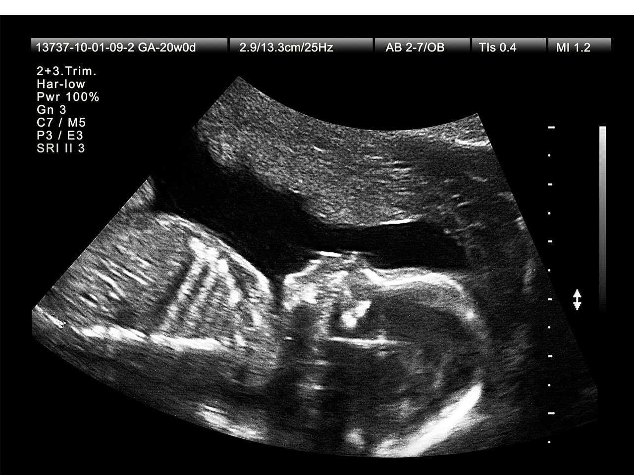
Fetal choroid plexus cysts and trisomy 18. The Ramzi method correctly predicts the fetus gender in 972 of the males and 975 of. An axial section of the brain including the thalami and the sphenoid bone wings should be obtained and magnified. Ultrasound is a widely used diagnostic procedure that takes pictures of the inside of the body using sound waves. Midway through your pregnancy between week 18 and week 22 a trained sonographer will perform a detailed anatomy scan called a level 2 ultrasound. Central Nervous System Diagnosis Of Fetal Abnormalities The 18 23 Fetal Abnormalities Diagnostic Medical Sonography Fetal.

The major fetal anomalies that can be recognized by ultrasound in the first trimester. Ultrasound images are captured in real-time so they can show the structure and movement of internal organs as well as blood flowing through blood vessels. - Assessment of basic anatomy after 11 week. A fetal ultrasound or sonogram is an imaging technique that uses high-frequency sound waves to produce images of a baby in the uterus. In general the main goal of a fetal ultrasound scan is to provide accurate information which will facilitate the delivery of optimized antenatal care with the best possible outcomes for mother and fetus. Pin On Baby Stuff.

- Assessment of basic anatomy after 11 week. Fetal Pole 55-6 weeks The fetal pole or developing embryo should be seen at 55-6 weeks gestational age by transvaginal ultrasound. Fetal heart sounds was described as early as 350 years ago and approximately 200 years ago mechanical stethoscopes such as the Pinard horn were introduced in clinical. Congenital hydrocephalus may be diagnosed by fetal ultrasound. The largest fetal organ the placenta undergoes rapid development over the course of pregnancy. Ultrasound Examination Of Fetal Anatomy 20 23 Weeks Venus Med.

Fetal Pole 55-6 weeks The fetal pole or developing embryo should be seen at 55-6 weeks gestational age by transvaginal ultrasound. Most of the time the condition is suspected during a physical exam following birth. With transvaginal ultrasound the fetal pole should be seen when it is 2-4mm in length. Typically the ultrasound is done halfway through the pregnancy. The second-trimester ultrasound is reassuring and fun to watch. Sound Waves Weekly Ultrasound Sonography Obstetric Ultrasound Medical Ultrasound.

Doppler ultrasonography is medical ultrasonography that employs the Doppler effect to generate imaging of the movement of tissues and body fluids usually blood and their relative velocity to the probeBy calculating the frequency shift of a particular sample volume for example flow in an artery or a jet of blood flow over a heart valve its speed and direction can be determined and. Fetal ultrasound has no known risks other than mild discomfort due to pressure from the transducer on your abdomen or in your vagina. Doppler ultrasonography is medical ultrasonography that employs the Doppler effect to generate imaging of the movement of tissues and body fluids usually blood and their relative velocity to the probeBy calculating the frequency shift of a particular sample volume for example flow in an artery or a jet of blood flow over a heart valve its speed and direction can be determined and. Medical Reviewers confirm the content is thorough and accurate. The major fetal anomalies that can be recognized by ultrasound in the first trimester. Pin On Ultrasound.

An NT is a special type of ultrasound using a very sensitive but safe machine. Aetna considers in-office and in-hospital antepartum fetal surveillance with non-stress tests NST contraction stress tests CST biophysical profile BPP modified BPP and umbilical artery and middle cerebral Doppler velocimetry medically necessary according to the American College of Obstetricians and Gynecologists ACOG Clinical Guideline on. Cardiotocography CTG is a technique used to monitor the fetal heartbeat and the uterine contractions during pregnancy and labourThe machine used to perform the monitoring is called a cardiotocograph. It is very likely that you will get a glimpse of what are now very obvious boy parts during this type of ultrasound. The major fetal anomalies that can be recognized by ultrasound in the first trimester. Pin On Sonography.

The major fetal anomalies that can be recognized by ultrasound in the first trimester. Anatomy Structure and Location. The Ramzi method correctly predicts the fetus gender in 972 of the males and 975 of. Anatomy of the Ventricular System. Typically the ultrasound is done halfway through the pregnancy. Nub Theory For Detecting Gender As Early As 10 Weeks Baby Gender Predictor Baby Gender Prediction Baby Gender Ultrasound.

CRL is measured as the largest dimension of embryo excluding the yolk sac and extremities. A fetal ultrasound or sonogram is an imaging technique that uses high-frequency sound waves to produce images of a baby in the uterus. Then he or she will locate the nuchal fold and measure its thickness on the screen. The largest fetal organ the placenta undergoes rapid development over the course of pregnancy. Crown rump length CRL is the length of the embryo or fetus from the top of its head to bottom of torso. Pin On Ultrasounds.

Predominantly fetal growth and organ maturation23. In these Guidelines we assume that the gestational age is known and has been determined as described above the pregnancy is singleton and the fetal anatomy is normal. Fetal Pole 55-6 weeks The fetal pole or developing embryo should be seen at 55-6 weeks gestational age by transvaginal ultrasound. With transvaginal ultrasound the fetal pole should be seen when it is 2-4mm in length. Ultrasound in the First Trimester 67 when compared to the abdominal transducers. Fetal Ultrasound With Subchorionic Hemorrhage At Seven Weeks Imaged With A Philips Envisor System This Is A Baby Ultrasound Ultrasound Fetal Baby Ultrasound.


